
SMILE™矫正近视伴高度散光术后角膜上皮厚度变化
作者:余克明 等
作者单位:中山大学中山眼科中心
发表杂志:Journal of Refractive Surgery
摘要

目的
研究SMILE™矫正中高度近视术后长期的角膜后表面高度变化。
方法
本前瞻性研究共计纳入接受SMILE™的40眼(23位患者),术前散光范围为-2.25至-4.50 D。除标准的眼科检查外,术前和术后也使用高分辨率谱域OCT测量9mm直径范围的角膜上皮厚度。分析残余散光度与术后上皮在术前平轴与陡轴方向上的厚度差异量之间的相关性。
结果
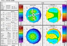
SMILE™术后6月,中央(2mm),旁中央(2-5 mm),中周部(5-7 mm)和周边区域(7-9 mm)角膜上皮厚度均有明显改变(P< .001)。其中,旁中央增厚最明显(9.75%),平均厚度最大(57.29 μm)。
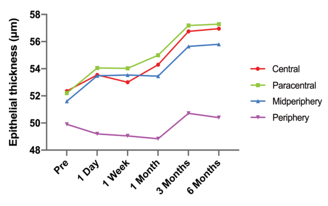
▲SMILE™术前与术后角膜中央(2mm),旁中央(2-5 mm),中周部(5-7 mm)和周边区域(7-9 mm)的平均上皮厚度。
此外,中周部的上皮在术前散光轴向方向有对称地区域性增厚。从术后1天至6月,中周部的上皮在术前平轴与陡轴方向的厚度差异持续增加。术后6月,该差异与残余散光度呈正相关,相关性有统计学意义(r = -0.034, P= .035)。
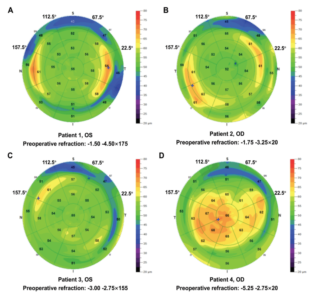
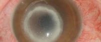
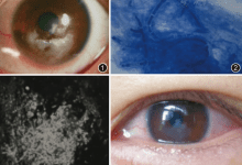
▲4位高度散光患者SMILE™术后角膜上皮厚度与其术前屈光度。
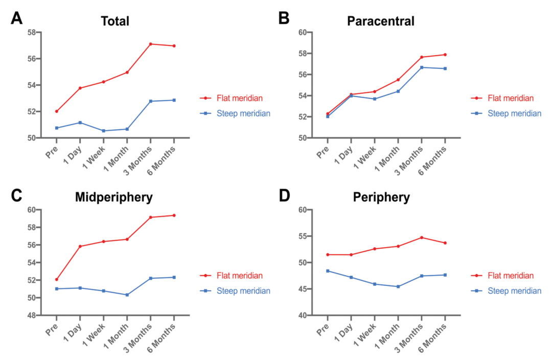
▲不同区域、不同术前术后时期,位于术前散光平轴与陡轴方向的平均上皮厚度。
结论
SMILE™矫正高度散光术后6月,9 mm直径范围内的角膜上皮厚度有明显增加。此外,中周部上皮在术前平轴与陡轴方向的增厚差异可能与SMILE™术后散光欠矫有关。
Corneal Epithelial Thickness Changes Following SMILE for Myopia With High Astigmatism
Keming Yu et al.
Abstract

Purpose
To evaluate the corneal epithelial thickness (CET) profile changes after small incision lenticule extraction (SMILE) surgery for myopic astigmatism correction of greater than 2.00 diopters (D).
Methods
This prospective observational study included 40 eyes (23 patients) treated with SMILE for myopia with cylinders of −2.25 to −4.50 D. Along with standard ophthalmic examinations, CET maps with a diameter of 9 mm were measured by high-resolution spectral-domain optical coherence tomography preoperatively and postoperatively. Correlations between the degree of residual astigmatism and the difference in CET values between preoperative flat and steep meridians were analyzed.
Results
The CET showed significant changes in the central (2 mm), paracentral (2 to 5 mm), midperipheral (5 to 7 mm), and peripheral (7 to 9 mm) zones 6 months after SMILE (P< .001). Among the regions, the CET in the paracentral zones displayed the largest increase (9.75%) with the highest average thickness (57.29 µm). Moreover, symmetrical regional epithelial thickening at the preoperative astigmatism axis was observed in the midperipheral zones. The difference in CET between preoperative flat and steep meridians in the mid-peripheral zones continuously increased from postoperative 1 day to 6 months. This difference was positively correlated with the residual cylinder errors at 6 months postoperatively (r= −0.334,P= .035).
Conclusions
The 9-mm diameter CET in eyes with high astigmatism significantly increased 6 months after SMILE. Additionally, the difference in CET between preoperative flat and steep meridians in midperipheral zones may be related to astigmatic undercorrection in SMILE.
本篇文章来源于微信公众号: SMILE屈光天地


 全飞秒
全飞秒 半飞秒
半飞秒 圆锥角膜
圆锥角膜 学术速递
学术速递