SMILE™术后帽与剩余基质厚度随时间的变化
作者:Markus Vicente Olsen 等
作者单位:挪威奥斯陆大学眼科
发表杂志:J Cataract Refract Surg.
Olsen, M. V., Lyngstadaas, A. V., Zhou, W., Stojanovic, A., Utheim, T. P., Wang, X., ... & Chen, X. (2020). Temporal redistribution of cap and residual stromal thickness after SMILE. Journal of Cataract & Refractive Surgery, 46(10), 1331-1338.
此前较少有研究报道SMILE™术后基质厚度的变化。并且,由于前40%的角膜弹力层拥有最强的生物力学特性,且胶原纤维的分布与深层角膜有所不同,因而可能导致术后重塑反应的不同。因此,本研究旨在探讨SMILE™术后角膜各个分层的厚度随时间的变化,以及厚度变化对屈光度或角膜地形图参数的影响,以进一步理解术后基质重塑对临床效果的影响。
摘要
目的
探究SMILE™术后角膜各个分层随时间的变化。
研究设计
回顾性。
方法
纳入接受SMILE™的51名患者的右眼。使用谱域OCT测量术后1天,1周,1月,3月,6月时的角膜上皮,帽,帽的基质部分,剩余基质床,总基质的厚度。分析各个分层厚度的变化,以及与等效球镜度变化、角膜前表面或后表面K值变化之间的相关性。
结果
从术后1天至术后6月,角膜各个分层的平均厚度都有增加:
角膜上皮:54.4 ± 4.0 µm至57.3 ± 5.2 µm
帽:137.1 ± 5.5 µm至140.3 ± 5.1 µm
帽的基质部分:82.7 ± 5.9 µm至82.8 ± 6.3 µm
剩余基质床:375.0 ± 40.8 µm至381.4 ± 30.6 µm
总基质厚度:457.6 ± 41.1 µm至462.1 ± 36.7 µm
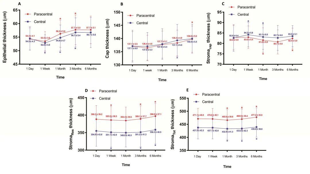
▲中央和旁中央的(A)上皮(B)帽(C)帽的基质部分(D)剩余基质床(E)总基质厚度随时间的变化。结果为平均值和标准差。*代表与前一个时间点的差异p < 0.05.
术后1月至6月之间,角膜中央前表面K值与帽的增厚,以及与帽的基质部分增厚的相关性有统计学意义。
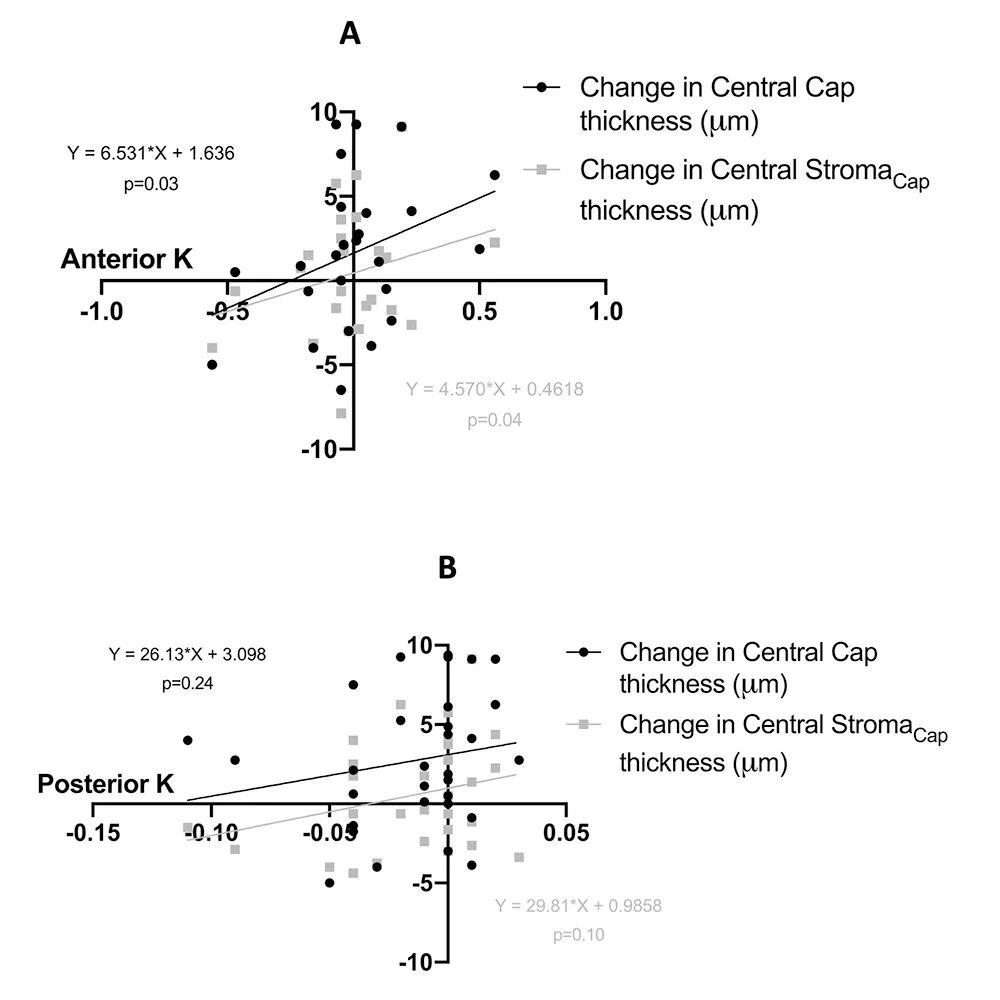
▲中央帽厚度,帽的基质部分厚度和(A)角膜前表面与(B)角膜后表面角膜曲率之间的相关性。
结论
SMILE™术后角膜前层(帽的基质部分)和后层(剩余基质床)基质的重塑反应不同。角膜前表面K值的变化,与中央帽或帽的基质部分之间有相关性。这可能是由于生物力学变化、组织重塑、愈合反应导致,或者是由于部分或全部上述因素的联合作用。
Temporal redistribution of cap and residual stromal thickness after SMILE
Markus Vicente Olsen et al.
Abstract
Purpose
To investigate corneal sublayer alterations during the postoperative period after small-incision lenticule extraction (SMILE).
Setting
Synslaser clinic, Oslo, Norway.
Study design
Retrospective.
Methods
Patients who underwent SMILE for treating myopia were included. The thicknesses of the corneal epithelium, cap, stromal part of the cap (StromaCap), residual stromal bed (StromaRes), and total stroma (StromaTot) were measured using spectral-domain optical coherence tomography at 1 day, 1 week, 1 month, 3 months, and 6 months postoperatively. Postoperative changes in the corneal sublayer thicknesses were analyzed and correlated with changes in spherical equivalence and anterior and posterior keratometry (K).
Results
The study was based on analyses of the right eyes of 51 patients. From 1 day to 6 months postoperatively, the corneal epithelium, cap, StromaCap, StromaRes, and StromaTot thicknesses increased from 54.4 ± 4.0 µm to 57.3 ± 5.2 µm; 137.1 ± 5.5 µm to 140.3 ± 5.1 µm; 82.7 ± 5.9 µm to 82.8 ± 6.3 µm; 375.0 ± 40.8 µm to 381.4 ± 30.6 µm; and 457.6 ± 41.1 µm to 462.1 ± 36.7 µm, respectively. Between 1 month and 6 months postoperatively, the increase in anterior K correlated significantly with the thickening of the cap (r = 0.37, P = .03) and the stromal component of the cap (r = 0.36, P = .04) within the central cornea.
Conclusions
The post-SMILE remodeling behavior between the anterior (StromaCap) and posterior (StromaRes) stroma were dissimilar. There was a significant correlation between changes in anterior K and the central cap and the stromal component of the cap. This might be because of biomechanical changes, tissue remodeling, and wound healing or a combination of some or all of the aforementioned processes.

点击“阅读原文”,可获取本文全文
星标置顶SMILE屈光天地
1.进入SMILE屈光天地公众号主页
2.点击右上角三个小点
3.点击“设为星标”

用三步
一眼找到SMILE屈光天地


本篇文章来源于微信公众号: SMILE屈光天地


 全飞秒
全飞秒 半飞秒
半飞秒 圆锥角膜
圆锥角膜 学术速递
学术速递