
强生Elita全飞秒SILK手术视频
简介强生Elita推出的全飞秒主要有以下优势:治疗范围:等效球镜在-1.00D至-12.00D,散光-6.0
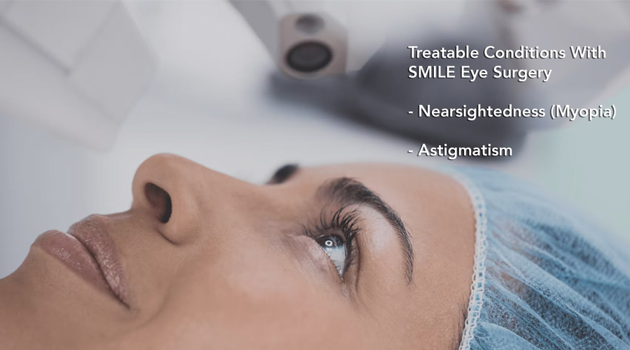
简介强生Elita推出的全飞秒主要有以下优势:治疗范围:等效球镜在-1.00D至-12.00D,散光-6.0

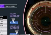
从目前媒体所能获得信息该机器主要有以下优势: 治疗范围:等效球镜在-1.00D至-12.00D,散光-6.00D。 低脉冲激光能量(亚微米级)和超快的激光重复率,达到1μm小光斑尺寸。 基质床更为光滑(SLIK技术),可以轻松分离透镜(从目...
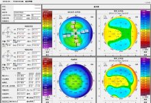
圆锥角膜的诊断需要分析断层地形图和/或角膜生物力学,还要结合患者年龄、眼睑情况、接触镜配戴、近视发展史等信息做出综合判断。环境不同,病有差异,专家观点仅供参考,他用无效。 本病例由云南大学附属医院梁刚主任分享 患者资料 患者女,24岁 近视...

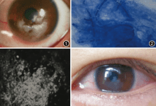
“全飞秒”术后出现真菌性角膜炎 一位41岁的女性因“右眼疼痛、发红3天”前来就诊。四天前,患者因双眼近视接受了双眼飞秒激光小切口角膜微透镜取出术(简称SMILE,又称全飞秒),术后患者局部使用左氧氟沙星和倍他米松滴眼液。专科检查:右眼最佳矫...
病例摘要 患者女性,23岁。因左眼视力下降、涩磨就诊。患者于2017年11月6日拟行近视眼矫正手术于解放军第一五九医院眼科就诊,术前视力右眼为0.01,左眼为0.01。双眼睑结膜轻度充血,可见散在滤泡和结石,球结膜无充血,角膜透明。2017...

 屈光说
屈光说