SMILE™与FS-LASIK矫正高度近视术后中央角膜基质减少量的可预测性:前瞻性、随机性、对侧眼研究
通讯作者:余克明 庄菁
通讯单位:中山大学中山眼科中心
发表杂志:J Refract Surg.
Luo Y, He S, Chen P, Yao H, He A, Li Y, Qiu J, Lan M, Zhuang J, Yu K. Predictability of Central Corneal Stromal Reduction After SMILE and FS-LASIK for High Myopia Correction: A Prospective Randomized Contralateral Eye Study. J Refract Surg. 2022 Feb;38(2):90-97. doi: 10.3928/1081597X-20211112-01. Epub 2022 Feb 1. PMID: 35156458.
背景:
凭借稳定、安全和可预测性良好的优势,SMILE™与FS-LASIK已成主流的角膜屈光手术技术,两种术式都有各自的优点。角膜屈光手术中,中央角膜厚度的消耗直接与预矫正屈光度相关,同时手术必须保留一定限度的角膜基质厚度,以防出现术后角膜扩张等并发症。因此,针对高度近视及角膜偏薄的患者,术式的选择和术前设计显得尤为重要。本研究对比了使用ZEISS VisuMax平台的SMILE™和使用Amaris 750S平台的FS-LASIK两种术式,在治疗高度近视术后患者中央角膜基质厚度的变化,为高度近视患者的屈光手术选择提供了较高水平循证医学证据。
摘 要
目的
比较SMILE™与FS-LASIK矫正高度近视术后中央角膜基质厚度减少量的可预测性。
设计
前瞻性、随机性、对侧眼研究。
方法
研究纳入42名高度近视患者患者,其中一只眼接受SMILE™(平均等效球镜度-8.339 ± 1.019 D),对侧眼接受FS-LASIK(平均等效球镜度-8.660 ± 1.197 D)。采用光谱域光学相干断层扫描技术(SD-OCT)测量角膜中央和上皮细胞的厚度,比较术前和术后的值,以确定中央基质的减少的量。
结果
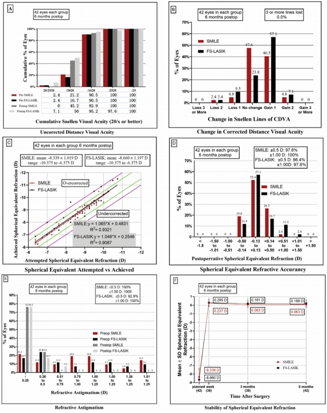
▲ SMILE™与FS-LASIK术后6个月随访时间内的屈光6联图(A:有效性;B:安全性;C:拟矫正与实际矫正SEQ散点图;D:可预测性;E:散光矫正;F:稳定性)
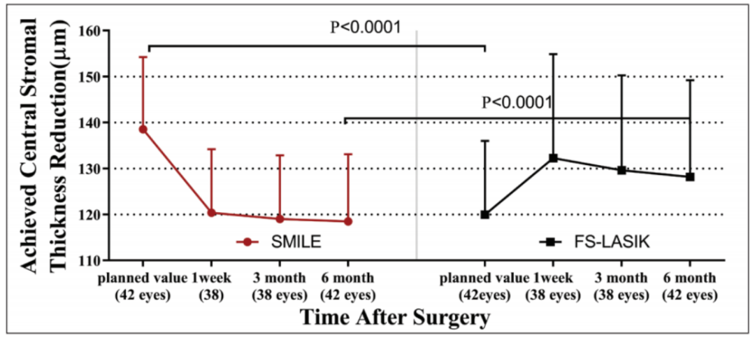
▲SMILE™与FS-LASIK术后6个月各随访时期内角膜基质厚度的改变量(红色点线为SMILE™,黑色方线为FS-LASIK)

▲SMILE™与FS-LASIK术后6个月实际中央角膜基质厚度改变量和预计改变量的散点图(A)和预计中央角膜基质厚度改变量与实际-预计改变量差值的散点图(B)
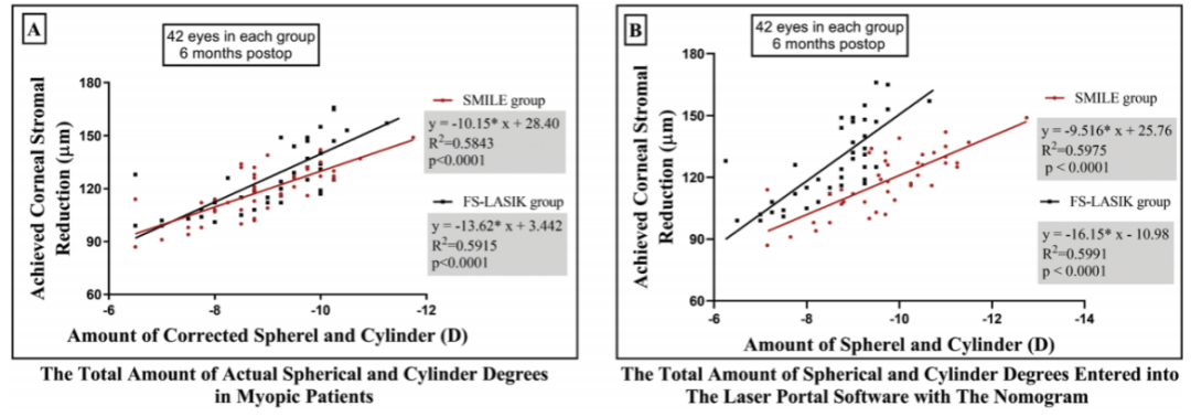
▲SMILE™与FS-LASIK组中,矫正屈光度和实际中央角膜基质厚度改变量的散点图(A为患者真实屈光度,即无nomogram调整,B为输机屈光度,包含nomogram调整,红色点线为SMILE™,黑色方线为FS-LASIK)

▲屈光可预测性(残余屈光度)与中央角膜基质厚度精确性(预计与实际角膜消耗厚度差异)的关系图(红色点线为SMILE™,黑色方线为FS-LASIK);相关性无统计学意义;
角膜基质厚度的变化:
术后6个月,SMILE™中央角膜基质厚度改变量被高估了20.05±5.92μm,FS-LASIK中央角膜基质厚度改变量被低估了8.21±8.14μm(p<0.0001)。在相同等效球镜的矫正中,SMILE™比FS-LASIK少消耗的中央角膜基质厚度平均为10.10±18.01μm(范围:1.90至18.29μm)。
角膜厚度消耗精确性与屈光矫正精确性:
术后6个月,不论是SMILE™还是FS-LASIK,均未发现预计与实际角膜基质消耗厚度的差异与屈光度的欠矫或过矫有关(p=0.9743 vs p=0.0777)。
结论
在高度近视患者的矫正中,SMILE™高估了实际角膜基质的消耗,FS-LASIK低估了实际角膜基质的消耗。在相同等效球镜的矫正中,SMILE™的角膜组织消耗量小于Amaris 750S屈光平台所完成的FS-LASIK。
Predictability of Central Corneal Stromal Reduction After SMILE and FS-LASIK for High Myopia Correction: A Prospective Randomized Contralateral Eye Study
Yiqi Luo, Shengyu He, Pei Chen, Huan Yao, Anqi He, Yan Li, Jin Qiu, Min Lan, Jing Zhuang, Keming Yu
Abstract
PURPOSE
To compare small incision lenticule extraction (SMILE) and femtosecond laser-assisted in situ keratomileusis (FS-LASIK) in terms of the predictability of central stromal thickness reduction in eyes with high myopia.
SETTING
State Key Laboratory of Ophthalmology, Zhongshan Ophthalmic Center, Sun Yat-sen University.
METHODS
In this prospective, randomized contralateral eye trial, 42 patients received SMILE in one eye and FS-LASIK (using the Amaris 750S excimer laser [SCHWIND eye-tech-solutions]) in the fellow eye for the correction of high myopia (manifest refraction spherical equivalent: < -6.00 diopters). Spectral-domain optical coherence tomography was used to measure the central corneal and epithelial thickness. Pre-operative and postoperative values were compared to determine the amount of central stromal reduction achieved.
RESULTS
At the 6-month follow-up visit, the amount of central stromal reduction was overestimated by 20.05 ± 5.92 µm in the SMILE group (P < .0001) and underestimated by 8.21 ± 8.14 µm in the FS-LASIK group (P < .0001). The mean actual central stromal reduction achieved with SMILE was significantly less than that achieved with FS-LASIK (10.10 ± 18.01 µm, range: 1.90 to 18.29 µm, P < .001). The discrepancy between the planned and achieved central corneal stromal reduction was not associated with refractive overcorrection or undercorrection in either the SMILE group or the FS-LASIK group (P = .9743 vs P = .0777).
CONCLUSIONS
In patients with high myopia, the laser software platform may underestimate and overestimate the amount of actual corneal reduction in eyes treated with FS-LASIK and SMILE, respectively. SMILE required less corneal stroma compared to FS-LASIK in the studied cohort using the Amaris 750S excimer laser when correcting a similar spherical equivalent refraction.
本篇文章来源于微信公众号: SMILE屈光天地










 全飞秒
全飞秒 半飞秒
半飞秒 圆锥角膜
圆锥角膜 学术速递
学术速递